The advent of fast and inexpensive computer workstations with high resolution color raster graphics has created the possibility of making attractive and sometimes insightful molecular images. However, the computer screen is two-dimensional and special techniques must be used to create the illusion of three dimensions. Moreover, three dimensions are not enough if we want to display continuously changing properties, like electrostatic potential or electron density.
Formally, the computer screen can be viewed as a plane (e.g., the XY plane of the cartesian coordinate system) and images of solid objects are projections of these objects onto the plane of the screen.
Early computer displays were capable of displaying lines and curves in a single color, and computer generated molecular representations were very similar to structural formulas drawn on paper. The addition of gradually varying intensity added a needed third dimension by making objects closer to the plane of the screen brighter. This technique is called depth-cueing, and it considerably enhances our perception of the three-dimensionality of objects. However, true stereovision results from the fact that each eye sees a slightly different picture because of slightly different visual angles. Computer stereovision is usually realized by a fast (e.g., 30Hz) successive display of images for the left and right eye which is synchronized with the operation of shutters in a special eye attachment or glasses. It frequently requires a special computer display or a screen attachment and such a system is called a stereoviewer. Early shutters where mechanical but lately polaroids or liquid crystal shutters are used.
The displays of the past could only show a limited palette of colors. This was adequate for coloring different atoms types but for continuously changing values, like electrostatic potential, it would only allow a display of a limited number of contours taken at discrete values. Modern computer displays allow an extensive number of colors (sometimes as many as a million) to be displayed. Colors combined with intensity variability allow a very perspicuous image of three dimensional objects. These capabilities are also used to paint molecular surfaces with a value of some property, like electrostatic potential or electron density.
To enhance perception of the three-dimensional shape of solid objects the technique of ray-tracing is frequently used. In this case the three-dimensional object (e.g., molecular surface) is treated as a reflective body placed behind the plane of the computer screen. One or more simulated light sources are placed somewhere in front of the computer screen (some may even be placed behind to illuminate the object) and a computer algorithm finds the reflections of the rays coming from these pseudo light sources and converts them into different intensities and colors. This gives a very powerful perception of shape and sculpture of the object surface.
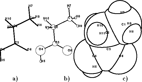
Figure: Molecular representations: a) stick model, b) ball-and-stick model,
c) space-filling model.
Molecules can be displayed in a variety of ways. The simplest is a stick
model of the molecule, a line drawing in which nuclei of bonded atoms
are connected by lines (Fig. 6.18a). The ball-and-stick
(Fig. 6.18b) models
are
stick drawings augmented with circles or balls whose radii are usually
taken as a fraction (e.g., 0.2) of the
corresponding van der Waals radii of atoms.
The van der Waals surface is another way of displaying a molecule. The surface
can be displayed either as dots (Fig. 6.14)
or a solid surface (Fig. 6.18c). Such
images are frequently called space filling or CPK models. In most cases molecular
images
are colored by atom type and their perception enhanced by depth
cueing and/or ray tracing techniques.
models. In most cases molecular
images
are colored by atom type and their perception enhanced by depth
cueing and/or ray tracing techniques.
Continuously changing values, like electrostatic potential, are inherently four-dimensional, i.e., they cannot be directly displayed even if a stereoviewer is used. Several approaches are possible in this case. The simplest is to choose a single plane and display the values as gradually changing color or intensity. By showing several such planes in succession (animation) or stacking them in such a way that they do not obscure each other, we can display fields or similar quantities at discrete planes in space. Another approach is to use contours. To produce a contour we have to choose a contouring level and then connect all points corresponding to this level. If the property to be contoured is continuous (i.e., changes gradually, without instantaneous jumps or drops) and bound (i.e., all its values are within a finite range) the contour is a curve (sometimes closed) in two dimensions (like you see on a map) or a surface in three dimensions.
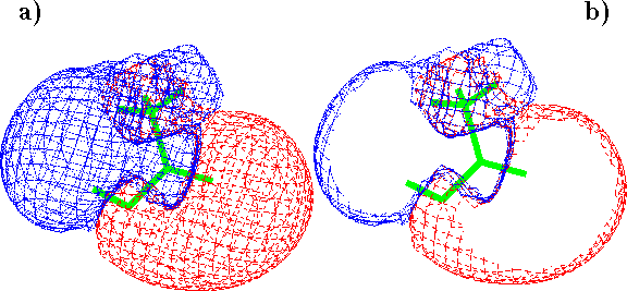
Figure: Electrostatic potential around acetic acid molecule contoured
at  and
and  levels: a) display of entire contours,
b) z-clipped contours
levels: a) display of entire contours,
b) z-clipped contours
Contours are usually created for several levels to show the variability of the contoured property. For two-dimensional cases it is easy to show different levels as different line styles. However, for three dimensions such contours would obscure each other. Transparent surfaces were only recently introduced to computer graphics and in most cases you will see three dimensional contours displayed as color dots or a system of intersecting lines called a wire frame or chicken wire. Their advantage is that you can display several of them simultaneously and they will not obscure themselves or the molecular image underneath. Color or line style is used to designate different contouring levels (see Fig. 6.19a).
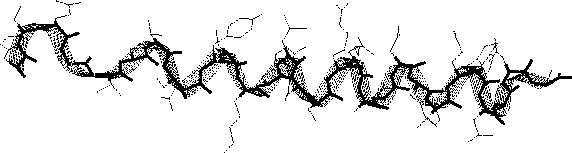
Figure 6.20: Display of ribbon and backbone for glucagon.
A computer display becomes difficult to follow if many three-dimensional objects are shown simultaneously. To help navigate among the different objects and see their relation, the technique of z-clipping is frequently used (Fig. 6.19b). In this method two planes parallel to the surface of the screen are used. Elements of the image sticking before the front plane and behind the back plane are clipped out, and only the portion in between clipping planes is displayed. This technique is used in conjunction with translating objects along the z-axis to allow selective viewing and close examination of interesting features of molecular models.
Displaying macromolecules is a challenge. The large number of atoms
makes the images very crowded and sometimes unviewable. To aid chemists
in exposing main structural features of the macromolecules the artificial
objects like ribbons,  -traces, etc. are displayed to highlight the
backbone of the macromolecule. The ribbon of
glucagon is represented in Fig. 6.20.
-traces, etc. are displayed to highlight the
backbone of the macromolecule. The ribbon of
glucagon is represented in Fig. 6.20.
Besides color, intensity, and shape, graphic systems also offer movement. Animation can be applied not only to dynamic properties of the system, like molecular dynamics trajectories, normal modes, etc., but can also be used to contrast objects against each another by displaying them in turns, by rotating them continuously with constant speed, or by rocking them. Sometimes blinking is used to highlight a fragment or molecule.
The computer graphics field is one of the most actively researched areas of computer science both from the psychological and engineering points of view. Methodological innovations are consistently incorporated into molecular modeling systems. It is however important not to get carried away with creating beautiful pictures. The graphics should illustrate and communicate chemical information and concepts in an easy to follow and clear way. Unfortunately, you can frequently see images overloaded with too many (however beautiful) details obscuring the underlying message.